Radiological imaging is a cornerstone of modern medicine, offering a window into the intricacies of human anatomy and pathology. Among the myriad of signs observed on X-ray films, the “steeple sign” is particularly noteworthy, often eliciting significant clinical interest. This phenomenon is predominantly associated with croup, a respiratory condition prevalent in pediatric populations. However, understanding the nuances of the steeple sign requires a deeper exploration into the underlying mechanisms of respiratory diseases and their radiological manifestations.
The Steeple Sign: An Overview
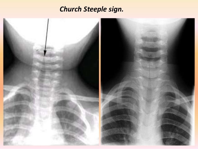
The steeple sign manifests as a distinct narrowing of the subglottic region of the larynx, resembling the shape of a church steeple on a frontal neck X-ray. This radiographic feature is characterized by an hourglass-like configuration, highlighting the roof of the trachea while emphasizing the constricted dimensions of the laryngeal space. Recognizing this sign is pivotal for practitioners, as it provides a visual correlate to respiratory distress stemming from upper airway obstruction, requiring prompt diagnosis and intervention.
Clinical Implications of the Steeple Sign
The primary condition associated with the steeple sign is croup, medically known as laryngotracheobronchitis. This inflammatory disease typically results from viral infections—most commonly, parainfluenza viruses—leading to the characteristic swelling of the larynx. Clinically, children with croup often present with a distinctive barky cough, stridor, and respiratory distress. The presence of the steeple sign on X-ray fortifies the clinical diagnosis, aiding healthcare practitioners in determining the severity of the condition and tailoring therapeutic interventions.
Pathophysiology Behind the Steeple Sign
The genesis of the steeple sign can be elucidated through a microscopic examination of the pathophysiological changes that occur during respiratory infections. In croup, viral agents trigger an immune response resulting in edema within the laryngeal framework. The consequent inflammation promotes narrowing at the level of the subglottic area. As the larynx becomes inflamed, it assumes a funnel shape, visually represented as the steeple sign on radiological evaluation. This anatomical alteration can lead not only to enhanced breathing difficulties but also to an increased risk of hypoxia in acute cases.
Differential Diagnosis: Beyond Croup
While croup is the prototypical condition associated with the steeple sign, other etiologies must be considered, particularly in atypical presentations or in populations outside the commonly affected pediatric demographic. Conditions such as epiglottitis—a bacterial infection leading to drastic swelling of the epiglottis—can occasionally result in similar X-ray findings. Anterior neck soft tissue imaging may render diagnostic confusion, necessitating a thorough assessment to differentiate these conditions. Other potential causes, albeit less common, include retropharyngeal abscesses and foreign body aspirations, each presenting unique challenges for clinical management.
Radiographic Techniques for Evaluating the Steeple Sign
To adequately visualize the steeple sign, practitioners often employ lateral and anteroposterior (AP) X-ray views of the neck. The lateral view is particularly beneficial, allowing for clear differentiation of the soft tissue contours surrounding the airway structures. The AP view, meanwhile, provides a broad perspective of potential mediastinal shifts that may accompany significant respiratory distress. In recent years, adjunct imaging modalities such as ultrasound and computed tomography (CT) have gained traction, providing visceral insight into complex cases where conventional X-ray may fall short.
Management Strategies for Croup
Management of croup hinges primarily on the severity of symptoms. Mild cases may be effectively managed with supportive care, including hydration, humidity, and close observation. Conversely, moderate to severe presentations warrant more aggressive pharmacological interventions. Systemic corticosteroids are the cornerstone of treatment to mitigate airway inflammation, while nebulized epinephrine may be utilized in acute exacerbations to counteract severe distress. In cases of impending respiratory failure, advanced airway interventions may become necessary, underscoring the importance of early recognition guided by radiological findings.
Conclusion: The Fascination Behind Radiological Signs
The steeple sign is more than a mere radiological hallmark; it encapsulates the intricate interplay of clinical prowess and imaging interpretation. Its identification not only aids in diagnosing croup but also provokes a broader conversation regarding the complexities of respiratory pathologies. As medical professionals continue to grapple with evolving respiratory challenges, the significance of such signs will undoubtedly endure, emphasizing the indispensable role of radiology in the realm of clinical practice. Through understanding and recognition, healthcare providers can offer timely and effective interventions, ensuring the health and safety of their patients.

Leave a comment