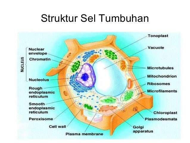
Histology is a fascinating field that allows us to explore the intricate architecture of tissues and cells within organisms. Within this realm, identifying distinct cell types is paramount for understanding both normal physiology and pathological conditions. But what if you had to discern which specific cell type is highlighted in a provided histological sample? This pursuit not only tests your observation skills but also challenges your understanding of cellular characteristics and functions. Let us embark on an exploration to unravel the components of cell identification in histology while posing the playful question: can you identify the cell type based on a visual fragment alone?
First and foremost, the anatomical orientation of the sample is crucial. When examining a histological slide, one’s first task is to discern the type of tissue represented. There are four primary types of tissues in the human body—epithelial, connective, muscular, and nervous. Each tissue has distinctive cell types with specific morphological features.
In an epithelial tissue, one might encounter squamous, cuboidal, or columnar cells, each offering a different architectural style. Squamous cells are flattened and have a scale-like appearance, designed for regulation and transport. Conversely, cuboidal cells are cube-shaped, adept at absorption and secretion, whereas columnar cells, taller and column-like, often contain goblet cells that secrete mucus. Understanding these characteristics paves the way to more focused identification.
Next, one must delve deeper into the histological intricacies of connective tissue. This category encompasses a diverse range of cell types, including fibroblasts, adipocytes, chondrocytes, and osteocytes, among others. Fibroblasts, for instance, are recognized by their elongated nuclei and abundant cytoplasm, playing a critical role in the formation of the extracellular matrix. In contrast, adipocytes, with their large lipid droplets, serve an entirely different function, primarily concerning energy storage. The presence of these characteristic features can decisively guide one toward accurate identification.
Then, we transition to muscular tissue, distinguished primarily by three types: skeletal, cardiac, and smooth muscle cells. Skeletal muscle fibers are multinucleated and exhibit striations, which are indicative of their contraction capabilities. Cardiac muscle cells, on the other hand, are interconnected via intercalated discs, allowing them to function as a cohesive unit—essential for the rhythmic contractions of the heart. Smooth muscle cells lack striations and have a spindle shape, contributing to involuntary movements in various organ systems. Recognizing these distinctions becomes imperative when identifying the highlighted cell type.
The Nervous tissue, composed of neurons and glial cells, presents its unique set of identification criteria. Neurons can be recognized by their distinct cell body, or soma, which houses the nucleus, long axons, and dendrites. Glial cells, meanwhile, serve various supportive roles and may be identified by their smaller size and varied shapes depending on their specific function within the nervous system.
Once the tissue type is determined, further examination of the specific cell features ensues. Various staining techniques are employed to highlight cellular structures. Hematoxylin and eosin (H&E) stain, the most commonly used, allows for differentiation between acidic and basic cellular components, providing a valuable visual contrast. With H&E sometimes obscuring fine details, differential staining methods like immunohistochemistry may be required to identify specific proteins or markers, adding another layer of detail to the identification process.
As one engages with these methodologies, one must not forget the significance of scale when observing histological samples. The resolution of the slides and the optical magnification used can profoundly influence your identification accuracy. A cell that might appear distinctly different at low magnification may reveal similarities when examined under higher power. Integrating this understanding into your analysis is a pivotal skill in the histological toolkit.
Additionally, recognizing the spatial organization of cells within a tissue section can provide clues to their identity. For instance, the arrangement of cells in a singular layer suggests epithelial tissue, while a more dispersed array indicates connective tissue. Understanding microenvironments is essential for identifying functional roles that specific cell types play within larger organ systems.
To add complexity to the cell identification challenge, one may consider the concept of pleomorphism, where cells of the same type can exhibit variability based on various factors such as age, disease state, or environmental conditions. This variability may necessitate a more nuanced approach to identification, moving beyond rigid categorizations to embrace a broader understanding of cellular biology.
In summary, the quest to identify a highlighted cell type in histology is an engaging challenge that melds keen observation with a deep understanding of biological principles. By systematically approaching the task—starting with overall tissue identification, moving into detailed cellular morphology, employing appropriate staining techniques, and considering cellular microenvironments—you arm yourself with the necessary tools for success. So, let the playfully posed question linger: are you ready to enhance your histological prowess and unveil the identity of the cell type you see before you? The journey of discovery awaits, promising rich rewards in both knowledge and expertise.
