In the realm of clinical neurology, the ability to assess the movement of extraocular muscles is indispensable. These muscles, which control eye movements in various directions, are integral to functioning in everyday life. The clinical test designed to evaluate these movements is characterized by its simplicity and effectiveness, yet it underpins a wide array of neurological insights. This article endeavors to examine the clinical test that assesses the movement of the extraocular muscles, elucidating the significance of this evaluation and its implications for broader neurological assessment.
Understanding Extraocular Muscles and Their Function
At the outset, it is essential to delineate the extraocular muscles. This group comprises six distinct muscles associated with each eye, specifically the superior rectus, inferior rectus, medial rectus, lateral rectus, and superior and inferior obliques. Collectively, these muscles govern the conjugate movements of the eyes, enabling coordination in gaze and visual tracking. Their optimal functionality is vital not only for vision but also for maintaining depth perception and spatial orientation. Dysfunctional movements can often manifest as diplopia, strabismus, or other ocular pathologies, highlighting the necessity of thorough clinical evaluation.
The Clinical Test: Evaluation of Extraocular Movements
The clinical test employed to assess the movement of the extraocular muscles is commonly referred to as the “extraocular movement test” or “six cardinal positions of gaze.” This examination involves the systematic observation of eye movements in six specific directions: up, down, left, right, and both oblique positions. Each movement is indicative of the integrity of cranial nerves III (oculomotor), IV (trochlear), and VI (abducens). The execution of this test is predominantly straightforward, yet the information it uncovers can be profound.
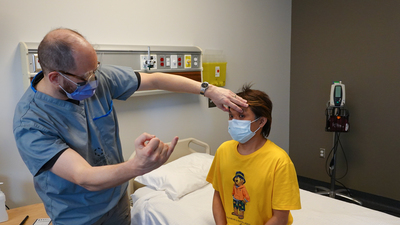
Conducting the Test: Methodology and Execution
To carry out the extraocular movement test, the clinician typically begins by ensuring that the patient is comfortably seated, maintaining a neutral head position. The practitioner then instructs the patient to follow a target—often a pen or finger—without moving their head. Observations are made as the clinician guides the target through the six cardinal positions. The clinician is vigilant for any irregularities or deviations in eye alignment and movement. The smoothness, range, and conjugacy of these movements are meticulously noted.
During the assessment, any jerky or disconjugate movements could imply underlying neurological issues, possibly involving the cranial nerves or muscular system. For instance, an inability to move the eye upward may suggest dysfunction of the oculomotor nerve, while an inability to adduct the eye could indicate a lesion affecting the abducens nerve.
Interpreting Findings: Clinical Significance
The clinical implications of the findings from this test are manifold. Anomalies in the movement of extraocular muscles can signify a plethora of conditions ranging from neurological disorders to systemic diseases. For instance, oculomotor nerve palsy commonly presents with a “down and out” position of the affected eye. In contrast, trochlear nerve palsy may result in vertical diplopia, especially when the patient is viewing downwards. Each of these presentations contributes to a differential diagnosis that could encompass vascular events, traumas, or even malignancies.
Deeper Neurological Connections
Moreover, the assessment of extraocular movements can unveil deeper neurological connections and highlight potential systemic issues. The conjugate eye movements are not isolated interactions; they are intricately linked to the vestibular system, which plays an essential role in balance and spatial orientation. Disruptions in eye movements may indicate broader vestibular or cerebellar dysfunction. This interrelation invites clinicians to contemplate not just the ocular implications but also the interconnectedness of neurological function.
Beyond the Test: A Broader Fascination
What captivates many clinicians and researchers in the field of neurology is the remarkable complexity of eye movement control. The precision of these movements belies an intricate choreography across numerous brain regions, including the brainstem, cerebellum, and cortical structures. This coordination reflects the brain’s ability to integrate sensory input from various modalities, further emphasizing the test’s role as a window into broader neurological function.
In examining the movement of extraocular muscles, clinicians are not only diagnosing isolated abnormalities; they are piecing together a puzzle that involves visual processing, motor function, and cognitive pathways. This holistic perspective fosters a deeper appreciation for the multifaceted nature of human physiology and the symbiotic relationships within various systems.
Conclusion
In conclusion, the clinical test of extraocular movements serves a vital function in the neurological examination process. By assessing the coordination and functionality of these muscles, healthcare professionals gain critical insights into cranial nerve integrity and potential underlying pathologies. The relatively straightforward execution of this test belies its complexity and importance within the broader context of neurological assessment. As clinicians engage with this examination, they are reminded of the intricate interconnectedness of bodily systems and the profound implications that arise from seemingly simple observations.

Leave a comment